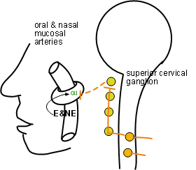
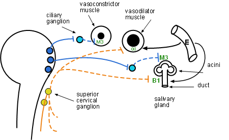
The main diagram illustrates several organs in the head: the vessels of the oral and nasal mucosae, the iris of the eye and salivary glands. p>

These arteries supply the lining of the mouth and nasal cavities. Arterial diameter is due to blood pressure pushing against the wall. If the tone of the encircling smooth muscle is increased the diameter can't expand as much thus restricting blood flow. If this tone is decreased then blood pressure can expand the diameter and increase blood flow.
Sympathetic nerves arising from the superior cervical ganglion innervate the encircling smooth muscle of these arteries. Norepinephrine activated alpha 1 receptors cause the cells to increase their tone and decrease blood flow. Epinephrine is especially potent in further increasing this tone resulting in additional vasoconstriction. The result is 'dry mouth' and 'ease of nasal air flow' that is part of the 'fight-or-flight' response characteristic of a high level of sympathetic activity. Notice there is no parasympathetic innervation at this site.

There are two smooth muscle arrangements in the iris. In the dilator muscle the fibers (elongated cells) are arranged radially around the pupil with the outer end of each cell anchored. There are alpha 1 receptors on these cells. In the constrictor muscle the fibers are arranged in concentric circles around the pupil and the 'head' and 'tail' of the fibers anchor on one another. There are muscarinic 3 receptors on these.
Parasympathetic neurons from the nearby ciliary ganglion (light blue circle) innervate the vasoconstrictor muscle. Acetylcholine activation of its M3 receptors causes constriction of these smooth muscle fibers and decreases pupil size.
Sympathetic neurons from the super cervical ganglion innervate the vasodilator muscle. Norepinephrine activation its alpha 1 receptors causes constriction. Due to their radial arrangement, and where each is anchored, the result is an increase in pupil size. Additional dilation can result from epinephrine diffusing from nearby capillaries.
The detailed physiological processes involved in saliva production are a 'hot topic' of research as of this writing. Salivary glands have flask-shaped acini of glandular cells that secrete amylase and/or mucus, along with fluid the consistency of blood plasma, into the hollow centers. The walls of the ducts have 'duct cells' that modify the composition of the passing saliva. Both divisions of the ANS are involved in the behavior of these glands.
Saliva is constantly produced because both divisions are always working simultaneously. The volume and consistency of the saliva depends on which division predominates at the given moment.
Acetylcholine activation of muscarinic 3 receptors on the glandular acini increases the secretion of saliva resulting in a high volume of watery saliva. This is characteristic of the 'rest-and-digest' response when parasympathetic activity dominates.
Norepinephrine activation of B1 receptors in certain duct cells results in low volume, viscous saliva. There is a good blood supply to these glands so that epinephrine decreases the volume even further under excessive sympathetic stimulation. The resulting 'dry mouth' is characteristic of the 'fight-or-flight' response at such times.